How AI Is Used in Radiology
If AI has one clear home base in medicine, it is radiology. More than three-quarters of all FDA-cleared AI medical devices are imaging tools, and the vast majority of those are built for reading X-rays, CT scans, and MRIs. This guide walks through what radiology AI actually does inside a real hospital workflow, not just what the marketing claims.
A radiology reading room is where most clinical AI in medicine is actually deployed today.
What Does AI Actually Do for a Radiologist?
Three things, mainly: it flags likely abnormalities so nothing gets missed in a busy queue, it reprioritizes urgent cases to the top of the worklist so the sickest patients are read first, and it drafts parts of the written report so radiologists spend less time on repetitive dictation. None of the three replaces the radiologist's judgment; all three change how their day is structured.
The scale here is genuinely large. As of March 2026, 1,163 of the FDA's 1,524 total cleared AI medical algorithms, 76.31%, are radiology tools, and the agency cleared 68 new radiology algorithms in just the first three months of the year. No other medical specialty is close. For the broader picture across all specialties, see our medical imaging overview.
How Does AI Reorder a Radiologist's Worklist?

AI tools in radiology typically highlight a suspicious region rather than issue a final verdict.
A busy radiology department can have hundreds of scans waiting to be read on a given day. Historically they were read in roughly the order they arrived. AI triage tools scan incoming images the moment they are captured and flag the ones showing signs of a likely emergency, such as a suspected brain bleed or a pulmonary embolism, moving them to the top of the queue automatically.
This is where some of the strongest published accuracy numbers in all of clinical AI show up. Intracranial hemorrhage detection tools have demonstrated 98.91% sensitivity and 99.83% specificity in prospective, multicenter studies. When a tool that reliable flags a scan, it genuinely shortens the time between a scan being taken and a radiologist looking at it, which for a bleeding brain can matter in minutes, not hours.
What Does the AI Actually Mark on a Scan?
| Tool type | What it flags | Reported accuracy |
|---|---|---|
| Aidoc body-CT (14 conditions) | Multiple acute findings across a single CT scan | 97% sensitivity / 98% specificity, FDA clearance Jan 2026 |
| Intracranial hemorrhage detection | Brain bleeds on head CT | 98.91% sensitivity / 99.83% specificity, prospective multicenter study |
| Mammography CAD tools | Suspicious masses and calcifications on breast imaging | Consistently in the high-80s to mid-90s percent range across cleared tools |
| Lung nodule detection | Small nodules on chest CT that could indicate early cancer | High sensitivity tools aimed at catching nodules easy to miss on a quick read |
In every case, the tool's output is a highlighted region or a flagged case, not a diagnosis written into the chart. The radiologist reviews the flagged area, agrees or disagrees with the AI, and writes the actual report. Some newer transparent-AI research, such as models trained on where radiologists' eyes actually look while reading a scan, is aimed at making that review process faster and easier to trust.
Does AI Also Help Write the Radiology Report?
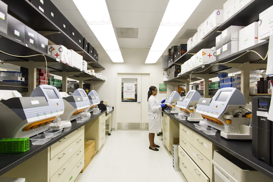
Behind the scenes, imaging AI tools are integrated directly into the software radiologists already use.
Increasingly, yes, and this is one of the fastest-growing uses of AI in the specialty. Report-drafting tools, sometimes bundled under names like Rad AI, listen to or read the radiologist's findings and generate a structured draft report in standard clinical language, which the radiologist then edits and signs. Reports suggest tools like this are now used in roughly half of US imaging practices, a workflow efficiency gain rather than a diagnostic one, since the underlying interpretation is still entirely the radiologist's.
This distinction matters for how you should read headlines about "AI writing your radiology report." The AI is drafting language around findings the radiologist has already made or confirmed. It is not independently interpreting your scan and generating conclusions on its own.
Why Did AI Take Off Here Before Everywhere Else?
Three structural reasons. First, medical images are already digital, standardized data, exactly the input format modern computer vision models are built to process, unlike, say, a doctor's handwritten notes or a patient's spoken history. Second, radiology has decades of labeled training data available in the form of past scans matched to confirmed diagnoses, which is the raw material any machine learning model needs at scale. Third, there is a persistent, well-documented radiologist workforce shortage in many countries, creating clear economic and clinical pressure to adopt tools that genuinely speed up the work.
Those same three conditions, digital input, abundant labeled data, and workforce pressure, are starting to show up in pathology too, which is why digital pathology is often called radiology's follow-on specialty for AI adoption.
Who Checks Whether These Tools Actually Work as Claimed?
FDA clearance is the first check, requiring a defined evidence bar before a tool reaches the market. But clearance summaries do not always disclose every detail of the pivotal study, which is why open-source, independently verifiable tools like Comp2Comp, a research platform for CT body composition analysis, matter: they let outside researchers examine exactly how an AI reaches its conclusions rather than trusting a closed, proprietary claim.
Ongoing, real-world monitoring matters just as much as the initial clearance study. A tool validated on one hospital's scanners and patient population can perform differently at another site with different equipment or demographics, which is why the FDA's newer "predetermined change control plan" framework increasingly requires manufacturers to monitor and report real-world performance after clearance, not just before it.
Should You Care Whether AI Reads Your Scan?
Practically, the level of care you receive should not change: a radiologist still reviews your images and is responsible for your report, whether or not AI flagged something first. What can change, and generally for the better, is speed. If your case is genuinely urgent, an AI triage tool may mean it gets looked at faster than it would have in a purely first-in, first-out queue. For the disease-detection angle beyond just imaging, see our disease detection guide.
How Does Radiology AI Fit Into the Rest of Healthcare AI?
Radiology's head start matters beyond radiology itself, because it is functioning as a proving ground for how AI gets validated, cleared, and trusted across the rest of medicine. The evidence standards, prospective trials, sensitivity and specificity reporting, human-in-the-loop deployment, that radiology AI has established are now being applied to newer categories like digital pathology and dermatology as those fields catch up.
If radiology is where AI in medicine is most mature, drug discovery is closer to the opposite end of that spectrum, promising but still mostly pre-clinical. Our guides on AI in drug discovery and AI-assisted medical research cover that earlier-stage frontier.
What Does an AI-Assisted Reading Day Actually Look Like?
A radiologist logs in and sees a worklist already reordered by AI triage, with a flagged possible hemorrhage sitting at the top instead of waiting in first-in-first-out order. They open that scan, and the AI has already drawn a highlighted box around the region it flagged. They review it, confirm or reject the AI's suggestion, and move to the next case, potentially with a draft report already generated for scans where findings are clear-cut.
What has not changed: the radiologist still looks at every scan, still makes the final call, and still signs the report under their own license. What has changed: the order they see cases in, how quickly the urgent ones get attention, and how much manual dictation time each report takes. That is the practical shape of "AI in radiology" today, a real workflow change wrapped around an unchanged clinical responsibility.
Frequently Asked Questions
What percentage of FDA-cleared AI is for radiology?
About 76%, or 1,163 of the FDA's 1,524 total cleared AI medical algorithms as of March 2026, making radiology by far the largest category of clinical AI.
Does AI replace radiologists?
No. Nearly every radiology AI tool is cleared and deployed as decision support: flagging findings, prioritizing urgent cases, or drafting reports, with a radiologist reviewing and finalizing every interpretation.
How accurate is radiology AI at detecting problems?
Reported figures vary by task but are generally strong for well-defined findings. Intracranial hemorrhage detection has shown 98.91% sensitivity in prospective studies, and a body-CT tool covering 14 conditions was cleared in 2026 at 97% sensitivity and 98% specificity.
What is AI report drafting in radiology?
It refers to tools that generate a structured written report based on findings the radiologist has already made or confirmed, saving dictation time. It is a workflow efficiency tool, not an independent diagnostic step.
More AI & Health Guides, Written Clearly
Browse our full library of AI and health explainers, updated as the tools and evidence change.
More from the creator
Explore two other projects from the same creator.